مجلات علمية


How does the heart work?
What is physiology?
Physiology is a science that aims to study the functions of the human body and how they work by studying the underlying chemistry and physics that intervene in the work of these functions, and depends on the various research studies of scientists that are conducted in the laboratory from studies on proteins and single cells to studies on the interaction of cells to form tissues, and organs, systems, inside the body1.
In general, physiology can be divided into 10 physiological organ systems as follows:
· Cardiovascular system.
· Digestive system.
· Endocrine system.
· Immune system.
· Muscular system.
· Nervous system.
· Renal apparatus.
· Reproductive system.
· Respiratory device.
· Skeletal device.
Cardiovascular system
The human heart is a finely-tuned instrument that serves the entire body. It is a strong organ around the size of a shut clench hand, and it sits in the chest, somewhat to one side of focus.
See how cardiovascular physiology is interlaced with other organ frameworks and how pathophysiology relates back to straightforward gross physiology. Heart physiology is one of the most significant bits of clinical information in medical services. The cardiovascular framework is continually adjusting to keep up with homeostasis in the body, explicitly to keep up with oxygen perfusion of tissues. The heart will adjust through numerous factors, for example, pulse, stroke volume, preload, afterload, diastole, and systole2.
The heart lives inside the pericardial sac and is situated in the mediastinal space inside the thoracic pit. The pericardial sac comprises of two melded layers: an external stringy container and an inward parietal pericardium fixed with a serous film. Between the pericardial sac and the heart is the pericardial depression, which is loaded up with greasing up serous liquid. The dividers of the heart are made out of an external epicardium, a thick myocardium, and an internal covering layer of endocardium3.
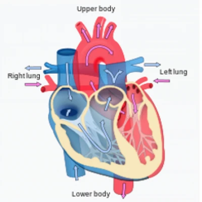
The human heart comprises of a couple of atria, which get blood and siphon it into a couple of ventricles, which siphon blood into the vessels. The right chamber gets foundational blood generally low in oxygen and siphons it into the right ventricle, which siphons it into the pneumonic circuit. Trade of oxygen and carbon dioxide happens in the lungs, and blood high in oxygen gets back to the left chamber, which siphons blood into the left ventricle, which thusly siphons blood into the aorta and the rest of the fundamental circuit. The septa are the segments that different the offices of the heart. They incorporate the interatrial septum, the interventricular septum, and the atrioventricular septum3.
Two of these openings are protected by the atrioventricular valves, the right tricuspid valve and the left mitral valve, which forestall the reverse of blood. Each is joined to chordae tendineae that stretch out to the papillary muscles, which are expansions of the myocardium, to forestall the valves from being blown once again into the atria. The aspiratory valve is situated at the foundation of the pneumonic trunk, and the left semilunar valve is situated at the foundation of the aorta. The right and left coronary conduits are quick to diverge the aorta and emerge from two of the three sinuses situated close to the foundation of the aorta and are by and large situated in the sulci. Heart veins equal the little cardiovascular conduits and for the most part channel into the coronary sinus3.
Pathway of blood move through the heart and lungs. Note that the aspiratory course (trunk) branches into left and right pneumonic conduits. There are generally four primary pneumonic veins that return blood from the lungs to the left chamber.
The conduction framework incorporates a few parts. The initial segment of the conduction framework is the sinoatrial hub. With no neural incitement, the sinoatrial hub musically starts driving forces 70 to 80 times each moment. Since it builds up the fundamental musicality of the heartbeat, it is known as the pacemaker of the heart. Different pieces of the conduction framework incorporate the atrioventricular hub, atrioventricular pack, group branches, and conduction myofibers. These parts facilitate the withdrawal and unwinding of the heart chambers4.
The cardiovascular cycle alludes to the substituting compression and unwinding of the myocardium in the dividers of the heart chambers, facilitated by the conduction framework, during one heartbeat. Systole is the compression period of the cardiovascular cycle, and diastole is the unwinding stage. At a typical pulse, one cardiovascular cycle goes on for 0.8 second5.
Diagrammatic conduction system of the heart
The sounds related with the heartbeat are because of vibrations in the tissues and blood brought about by conclusion of the valves. Unusual heart sounds are called mumbles.
The sinoatrial hub, acting alone, produces a steady musical pulse. Directing components are dependent on the atrioventricular hub to increment or diminishing the pulse to change heart yield to meet the changing requirements of the body. Most changes in the pulse are intervened through the heart place in the medulla oblongata of the mind. The middle has both thoughtful and parasympathetic parts that change the pulse to meet the changing requirements of the body.
Fringe factors like feelings, particle focuses, and internal heat level might influence pulse. These are normally interceded through the heart place.
Blood is the liquid of life, moving oxygen from the lungs to body tissue and carbon dioxide from body tissue to the lungs. Blood is the liquid of development, shipping sustenance from absorption and chemicals from organs all through the body. Blood is the liquid of wellbeing, shipping sickness battling substances to the tissue and waste to the kidneys. Since it contains living cells, blood is alive. Red platelets and white platelets are liable for supporting and purging the body.
Johannes Purkinje (1787–1869) was a productive Czech anatomist and physiologist6. He set up the main Department of Physiology on the planet in 1839 in Prussia. He was quick to utilize a microtome to acquire slim tissue areas for minute assessment and was quick to portray sweat organs. He presented the expressions "plasma" and "cellular material." He made significant commitments to the investigation of vision, including disclosure of the Purkinje impact, the Purkinje shift, and Purkinje pictures. He found two sorts of Purkinje cells: huge neurons in the cerebellum and, exceptionally compelling to heart electrophysiologists, huge cardiovascular muscle filaments specific for fast conduction along the endocardium of the ventricles7.
Purkinje filaments contrast in various ways from working myocardial cells. The relative sizes of Purkinje and myocardial cells are species-dependent. As a general rule, Purkinje cells are bigger than myocardial cells and are discernable histologically. Purkinje cells need cross over tubules that are available in myocardial cells. Because of their essential job in quick conduction of the electrical drive, Purkinje cells have less myofibrils than myocardial cells. Thus, infinitesimally they seem lighter than myocardial cells. The measure of glycogen in Purkinje fiber is a lot higher than that in myocardial cells. The glycogen can be utilized anaerobically which might make Purkinje cells more protection from hypoxia than working myocardial cells, albeit some proof recommends that the inverse is true6.
Purkinje fiber just beneath the endocardium.
The heart is a fundamental, incredible organ that continually siphons oxygen and supplements around the body.
In the event that an individual is brought into the world with inherent coronary illness, or then again if harm happens because of sickness or different components, the heart's capacity might decrease, and this can prompt hazardous intricacies, like cardiovascular breakdown.
In the event that the heart stops, an individual can't make due for long. Remaining dynamic and keeping a restorative eating routine are two different ways to ensure the heart.
References
1. https://www.medicalnewstoday.com/articles/248791
2. Oberman R, Bhardwaj A. Physiology, Cardiac. 2021 Jul 21. In: StatPearls [Internet]. Treasure Island (FL): StatPearls Publishing; 2021 Jan–. PMID: 30252345.
3. Rehman I, Nassereddin A, Rehman A. Anatomy, Thorax, Pericardium. [Updated 2021 Jul 27]. In: StatPearls [Internet]. Treasure Island (FL): StatPearls Publishing; 2021 Jan-. Available from: https://www.ncbi.nlm.nih.gov/books/NBK482256/
4. Kashou AH, Basit H, Chhabra L. Physiology, Sinoatrial Node. [Updated 2020 Oct 6]. In: StatPearls [Internet]. Treasure Island (FL): StatPearls Publishing; 2021 Jan-. Available from: https://www.ncbi.nlm.nih.gov/books/NBK459238/
- Grøndal N, Bramsen MB, Thomsen MD, Rasmussen CB, Lindholt JS. The cardiac cycle is a major contributor to variability in size measurements of abdominal aortic aneurysms by ultrasound. Eur J Vasc Endovasc Surg. 2012 Jan;43(1):30-3. doi: 10.1016/j.ejvs.2011.09.025. Epub 2011 Oct 21. PMID: 22018525.
- Jones, Daniel (2011). Roach, Peter; Setter, Jane; Esling, John (eds.). Cambridge English Pronouncing Dictionary(18th ed.). Cambridge University Press. ISBN 978-0-521-15255-6.
- Robinson V. Johannes Evangelista Purkinje. Scientific Monthly. 1929;XXXIX(3):217–229.